Obesity triggers high blood pressure, high blood sugar, and lipid abnormalities, leading on to such serious diseases as arteriosclerosis, heart attack, stroke, dementia, and diabetes. It would thus be fair to say that obesity truly is the prime culprit in lifestyle diseases. Remedying obesity is also a pressing issue in order to extend healthy life expectancy and curb medical expenses, but there was hitherto no effective means of combatting it. However, scientists have now shed light for the first time on the mechanism via which the numerous mitochondria in brown adipocytes mediate the activation of this type of adipose tissue and thereby encourage fat burning. Hopes are growing that this will be the trump card in tackling obesity.
Special Feature 1 – The Impact of Mitochondria Activating brown adipocytes to promote fat burning
text by Toshiko Mogi

Masakazu Fujii
Co-Investigator, Department of Clinical Chemistry and Laboratory Medicine, Graduate School of Medical Sciences, Kyushu University
Joined the diabetes laboratory at Kyushu University’s Third Department of Internal Medicine after graduating from the University of Toyama’s Faculty of Medicine in 1998. Between 2013 and 2016, he was involved in basic research into vascular biology at Emory University in the U.S. After returning to Japan, he was appointed assistant professor in the Department of Clinical Chemistry and Laboratory Medicine at Kyushu University’s Graduate School of Medical Sciences, subsequently serving as a part-time lecturer before taking up his current post. Following a period serving as deputy director of Fukuoka Prefectural Social Insurance Medical Association Inatsuki Hospital, he took up the post of diabetologist in the Department of Diabetology at Itoshima Medical Association Hospital in 2024. With a primary focus on diabetes, he is involved in the treatment of lifestyle diseases from both the research and clinical perspectives.
A team of researchers has discovered the self-activation and fat-burning mechanisms of brown adipocytes mediated by mitochondria; research aimed at drug discovery is continuing. We spoke to Dr. Masakazu Fujii from the research team to find out more.
He begins with a question: “Have you heard of the metabolic domino effect?”
The metabolic domino effect refers to a situation in which obesity triggers metabolic syndrome in the form of hypertension, hyperglycemia, and dyslipidemia, which then leads to such serious diseases as arteriosclerosis, myocardial infarction, stroke, dementia, diabetes, and metabolic dysfunction-associated steatohepatitis (MASH), in a manner similar to a row of dominoes falling.
“The first domino that sets the rest falling is obesity.”
Preventing a buildup of visceral fat is most effective
Obesity is broadly classified into two categories: primary obesity, which is caused by lifestyle; and secondary obesity, which is caused by diseases such as Cushing’s syndrome and hypothyroidism. Primary obesity is often the type that serves as the first domino.
Based on his encounters with numerous patients in his work as a diabetologist, Fujii identifies two key culprits in obesity: overeating and physical inactivity. If the amount of energy burned is less than the amount of energy consumed, the excess energy will be stored by the body as fat and the individual will remain obese.
The excess fat stored by the body is divided into two types: subcutaneous fat and visceral fat. Subcutaneous fat is the kind of fat that accumulates around the flanks and that can be pinched between one’s fingers, whereas visceral fat builds up around the stomach, intestines, and other internal organs in the abdominal cavity. It is the latter, visceral fat, that poses a high risk of triggering the metabolic domino effect.
“Preventing a buildup of visceral fat—the first domino in the chain—is the most effective way to keep the metabolic domino effect at bay,” Fujii says. “And the cheapest and most effective means of doing that is weight loss.”
In other words, one needs to improve one’s lifestyle by remedying overeating and taking moderate exercise in order to ensure one’s energy balance does not go into surplus. However, this is difficult. In many cases, people are able to lose weight temporarily through treatment focused on such aspects as diet and exercise. But this weight loss does not continue.
“Oral and injectable medications aimed at treating obesity are the focus of tireless R&D and commercialization efforts around the world. They’re also used in real-life clinical settings in Japan, where they’re covered by insurance for the treatment of obesity. However, it’s essential to have a full understanding of the appropriate usage and side effects of each individual drug,” Fujii continues. “For cases of severe obesity, where people have great difficulty following therapeutic diet and exercise regimes, there are also surgical treatments that remedy obesity by reducing the size of the stomach in order to reduce calorie intake.”
In 1992, the drug mazindol was made eligible for insurance coverage as a drug to treat obesity. It acts on the hypothalamus to suppress appetite, but great care must be taken with regard to safety, dependence, and side effects, as its pharmacological properties are similar to those of stimulants.
The leading drugs to treat obesity that have been made eligible for insurance coverage in recent years are semaglutide in 2023 and tirzepatide in 2025. Both were already approved as drugs to treat type 2 diabetes, but they are now covered by insurance as drugs to treat obesity in cases where an individual is medically diagnosed with obesity and the prescribing institution meets the required criteria. In addition to their action in lowering blood glucose levels, these drugs act on the brain’s appetite center to suppress appetite and sustain feelings of satiety, which lead to weight loss. However, side effects primarily affecting the gastrointestinal system have been observed, including nausea, vomiting, constipation, diarrhea, abdominal pain, and abdominal bloating, and there have even been reports of serious side effects such as acute pancreatitis and cholecystitis.
“Consequently, there has, until now, been no perfect means of treating obesity.”
According to the Ministry of Health, Labour and Welfare’s National Health and Nutrition Survey (2023), 31.5% of men and 21.1% of women aged 20 or over in Japan are obese. The highest proportion of obese men is found among those in their 60s, at 35%, and the proportion tends to increase with age. In addition, the proportion of obese people increased between 2013 and 2019, with no subsequent increase or decrease observed. Considering the current state of society, remedying obesity is also becoming a pressing issue in order to extend healthy life expectancy and curb medical expenses.
“The research we undertook marks the beginning of efforts to improve obesity treatment,” Fujii says. “We unraveled the mechanism promoting fat burning that is triggered when the mitochondria in brown adipocytes are activated.”
Brown adipocytes contain a large number of mitochondria
Let us now look at fat cells. Fat cells are divided into two types: white adipocytes and brown adipocytes (Figure 1).
Figure 1. The two types of adipose tissueOn the left is white adipose tissue, while on the right is brown adipose tissue. Brown adipocytes contain a very large number of mitochondria and produce heat.
“Brown adipose tissue looks brown even to the naked eye, because the cells contain an exceptionally large number of mitochondria,” Fujii explains. “And mitochondria are known to generate heat within cells.”
For many years, scientists thought the purpose of brown adipocytes was to maintain body temperature in newborn infants.
“Brown adipocytes generate heat in order to ensure that, when babies first emerge from the warmth of the womb, their body temperature remains constant after their birth, despite their abrupt emergence into a cold environment.”
Upon exposure to cold stimulus, the brain issues an order via the sympathetic nervous system, instructing the body to produce heat to maintain body temperature. In response, the sympathetic nerve endings release noradrenaline. When this noradrenaline stimulates brown adipocytes, stored fat is broken down, producing fatty acids. These fatty acids serve as a source of energy within mitochondria, resulting in the production of heat via the heat-producing protein UCP-1. It is thought that this is how body temperature is maintained.
Scientists used to think that brown adipocytes existed only during infancy and disappeared as the body grew. However, in 2009, scientists discovered that brown adipocytes also exist in adults and confirmed that it is primarily distributed around the clavicles, neck, and scapular region.
“From that point, scientists began to focus on the activation of brown adipocytes, which burn fat to produce heat, and research aimed at helping to remedy obesity has been flourishing in Japan and overseas ever since.”
However, no drugs to treat obesity have as yet been successfully developed for clinical use as a result of this research. Amid this situation, the Department of Clinical Chemistry and Laboratory Medicine at Kyushu University’s Graduate School of Medical Sciences embarked in 2019 on a study aimed at combatting obesity by activating brown adipocytes via mitochondria.
“The Department of Clinical Chemistry and Laboratory Medicine conducted a collaborative study with the Department of Cardiovascular Medicine in 2005, in which transgenic mice overexpressing the protein mitochondrial transcription factor A (TFAM) were generated. The article they published reported that, even after myocardial infarction, these TFAM-overexpressing mice (TgTg mice) demonstrated better left ventricular function than wild-type mice that had not been genetically modified (WT mice), and also had a higher survival rate,” Fujii explains.
This study revealed that mitochondrial function is elevated in TgTg mice. Interestingly, the results of the study also showed that the TgTg mice maintained a lower body weight than WT mice, and also lived longer.
“However, since 2005, nobody had investigated the mechanism that would explain why the TgTg mice weighed less and lived longer,” he continues. “After returning home from my studies in the U.S., I was involved in obesity research in the diabetes laboratory, so I became part of a new collaborative research team and began working to shed light on the anti-obesity mechanism in TgTg mice.”
The factor that activates brown adipocytes
The key to solving this puzzle is TFAM.
“This protein coils around mitochondrial DNA and is arranged in such a way as to maintain its structure,” Fujii explains. “Consequently, scientists believe that it is involved in the transcription and replication of mitochondrial DNA. However, we also believe it has a variety of other functions, but these have yet to be fully elucidated.”
The study used WT and TgTg mice, with TFAM expression levels in the TgTg mice approximately 2.5 times higher than in the WT mice.
When the research team examined differences in body weights after six months on a normal diet, the WT mice weighed 40.4 g, whereas the TgTg mice weighed markedly less, at 26 g, demonstrating resistance to obesity.
“We then fed the TgTg mice and WT mice a high-fat diet (in which fat-derived calories accounted for about 60% of total calorie intake) for eight weeks, and found that while the the WT mice gained weight and became obese, no weight increase was observed in the TgTg mice,” Fujii says (Figure 2).
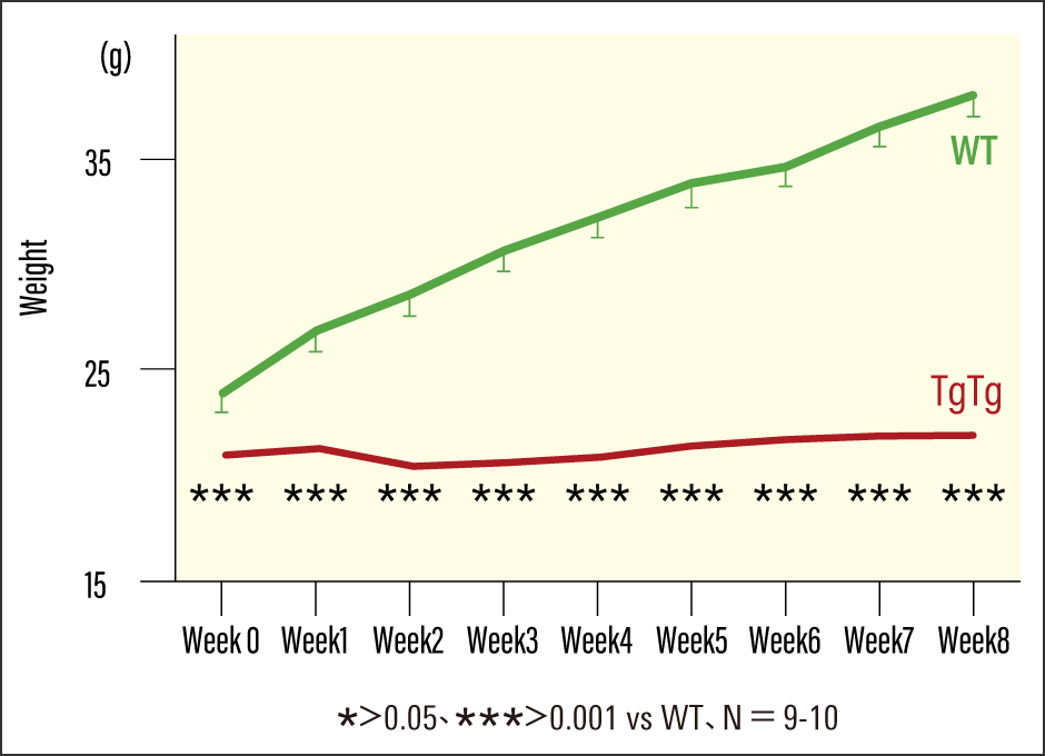
Figure 2. Changes in the body weights of TgTg mice and WT miceWhen fed a high-fat diet continuously for eight weeks, the WT mice gained weight, but there was no change in the weight of the TgTg mice.
When the researchers examined the adipose tissue under a microscope, they found that whereas the white adipocytes of WT mice were enlarged, there were no such changes in the TgTg mice.
“We looked at their livers and found that those of WT mice appeared whitish, due to the accumulation of fat, whereas those of TgTg mice were reddish color, as no fat had accumulated.”
The research team next performed an experiment in which they harvested brown adipocytes from both TgTg and WT mice, cultured and expanded them, and transplanted them adjacent to the brown adipose tissue of the WT recipient mice. Under a high-fat-diet, the TgTg-t mice that had received transplants of TgTg-derived cells showed marked suppression of weight gain compared with the WT-t mice that had received WT-derived cell transplants (Figure 3). This suggests that the brown adipocytes from the TgTg mice activated brown adipocyte tissue in the WT mice.
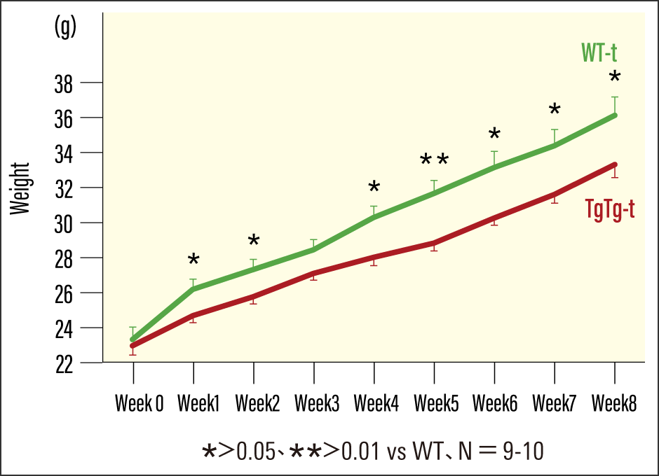
Figure 3. Changes in the body weights of TgTg-t mice and WT-t miceThe research team harvested brown adipocytes from both TgTg and WT mice, cultured and expanded them, and transplanted them into a location adjacent to the brown adipose tissue of the WT mice. When fed a high-fat diet for eight weeks, the TgTg-t mice that received transplants of TgTg-derived cells exhibited an anti-obesity effect.
“Based on the results of this experiment, it was hypothesized that the high TFAM-expressing brown adipocytes secrete a humoral factor that promotes brown adipocyte activation in an autocrine (where a secreted factor acts on the secreting cell itself) or paracrine (where a secreted factor acts on neighboring cells) manner,” Fujii explains. “Based on this hypothesis, we undertook further experiments.”
The research team carried out a special cell culture technique called co-culture: they divided the space between the TgTg mouse-derived and WT mouse-derived brown adipocytes with a special membrane through which only substances of a certain size could pass, allowing the culture media to communicate across the membrane. They found that activation of the cells occurred not only in brown adipocytes derived from the TgTg mice, as expected, but also in brown adipocytes derived from the WT mice.
Ucp-1 and Pgc-1α are markers known to be indicative of brown adipocyte activation. Activated in response to exposure to the cold and other stimuli, brown adipocytes produce heat, which causes the level of Ucp-1 expressed to rise. In addition, Pgc-1α boosts mitochondrial function, which promotes Ucp-1 expression. Increases in the expression of both (at both the gene and protein levels) were observed in co-cultured WT mouse-derived cells, too.
What, then, was the substance capable of passing through this special membrane? Subsequent detailed analyses identified it as exosomes (extracellular vesicles). Exosomes excessively secreted from TgTg mouse-derived cells were shown to pass through the membrane and act on WT mouse-derived cells, thereby inducing their activation.
“From these findings, we confirmed that in brown adipocytes derived from TgTg mice, elevated mitochondrial function increases extracellular secretion of exosomes, which reach WT mouse-derived cells via the culture medium and contribute to their activation.”
Physiologically present in the body, exosomes are capsule-like substances secreted by cells; they carry proteins and other components, as well as RNA and other information about cells. By transporting these components and information around the body, exosomes help cells to work together and contribute to cellular repair, regeneration, and activation.
The research team continued to carry out various experiments thereafter. Ultimately, this is what they discovered: in brown adipocytes that express a high level of TFAM—mitochondrial transcription factor A—mitochondrial function increases, promoting the secretion of exosomes. The exosomes secreted in greater numbers are taken up by the secreting cells themselves as well as by neighboring cells. The team became the first in the world to shed light on the mechanism by which this activation of brown adipocytes promotes fat burning (thermogenesis) and exerts an anti-obesity effect (Figure 4).
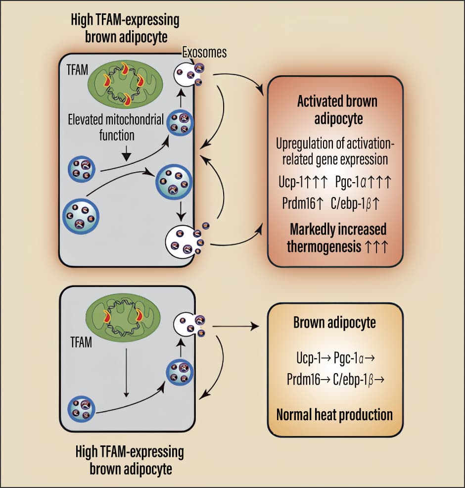
Figure 4. Mechanism involved in the activation of brown adipocytes and fat burningThe image at the top shows the mechanism discovered through the study, while the one at the bottom shows the normal mechanism. In high TFAM-expressing brown adipocytes, mitochondrial function is elevated, promoting the secretion of exosomes. These are taken up by the secreting cells themselves and by surrounding cells, causing a rise in the expression of genes and proteins related to brown adipocyte activation. This self-activation mechanism brings about a sustained increase in thermogenesis, thereby exerting a powerful anti-obesity effect.
Progress toward drug development
The findings from this study attracted a great deal of attention, because they offered hope that they would lead to the development of a drug providing a fundamental treatment for obesity by burning fat itself, rather than conventional drugs that act by suppressing appetite.
“We’re continuing our research with a view to clinical applications—specifically, drug discovery,” Fujii says. “Informed by our basic research to date, we’re exploring several avenues for drug development based on our exosome research, including (1) administering human adipocyte-derived exosomes themselves; (2) exploring drugs that enhance mitochondrial function and thereby induce exosome secretion; and (3) identifying substances within the exosomes that demonstrate anti-obesity effects and pursuing drug development accordingly. I believe that this may enable us to develop an unprecedented therapeutic agent for obesity that can promote fat burning within patients.”
Genetically modified TgTg mice were used to elucidate the anti-obesity mechanism mediated by elevated mitochondrial function. However, now that the team is moving toward translational research, they are focusing on exosomes derived from the brown adipocytes of physiologically intact WT mice without genetic modification.
“Currently, we’re isolating exosomes—which may be regarded as anti-obesity factors—from the brown adipocytes of WT mice and evaluating their cellular effects,” Fujii explains. “We also have established an immortalized cell line (a cell line capable of proliferating indefinitely) to create a stable system for exosome production.”
They have also established a method for purifying and concentrating large quantities of exosomes.
“Exosomes contain a wide range of functional proteins,” he continues. “We need to ascertain which of the substances they contain exhibit an anti-obesity effect. Our research team is working on that question right now, as well.”
They have already mapped out a solid blueprint for clinical studies. The goal is to stop the metabolic domino effect in its tracks.
“If we can stop the first domino from toppling the rest, we could well be able to prevent or remedy a variety of diseases that start from obesity,” Fujii says.
Expectations for the research findings are rising.
(Figures courtesy of Masakazu Fujii)