Measuring just 5 µm in internal diameter, the ultra-fine blood vessels called capillaries are known to play an important role in supplying oxygen and nutrients to cells and tissues. As we age, our capillaries become even finer and sparser. Once they enter this state, becoming what are called “ghost vessels”, the blood flow through the capillaries declines and they become unable to transport oxygen and nutrients. As a result, the cells and tissues around the ghost vessels gradually become damaged and their function deteriorates. The latest research has identified a relationship between these ghost vessels and a variety of diseases, including lifestyle-related diseases such as diabetes and hypertension.
Special Feature 1 – Understanding Blood Vessels Ghost vessel formation in capillaries is closely associated with lifestyle-related diseases
composition by Takakazu Kawasaki

Nobuyuki Takakura
Professor, Department of Signal Transduction, Research Institute for Microbial Diseases, Osaka University
Graduated from the Faculty of Medicine at Mie University in 1988 and entered clinical practice as a hematologist. He subsequently embarked on basic research with the aim of developing groundbreaking cancer remedies and tissue regeneration therapies. In 1997, he successfully completed the doctoral program at Kyoto University's Graduate School of Medicine and was awarded a Ph.D. in medicine. That same year, he became a research associate at the School of Medicine and the Institute of Molecular Embryology and Genetics, Kumamoto University. In 1999, he became an assistant professor at the same school, taking on the role of associate professor at the institute the following year. In 2001, he became a professor at the Cancer Research Institute, Kanazawa University. He has held his current post since 2006.
It has been discovered that deteriorating capillary function exacerbates lifestyle-related diseases such as diabetes and hypertension, and can also trigger dementia, osteoporosis, liver dysfunction, and renal disease. It is also closely related to cancer and aging.
The main function of our blood vessels is to transport oxygen and nutrients to the 37 trillion (or, in some theories, 60 trillion) human cells in an instant. Accordingly, our bodies are densely packed with blood vessels. Computer calculations have estimated that if all the blood vessels in a single human body were placed end to end, they would stretch for a distance of approximately 100,000 km—long enough to circle the earth about two and a half times. Capillaries account for more than 95% of this figure.
Blood flows from arteries to arterioles, capillaries, venules, and finally to veins. Classifying blood vessels by function and structure, we can divide them into three categories: arteries, arterioles, and veins; venules; and capillaries.
Carrying out carbon dioxide and supplying nutrients
A key characteristic of arteries, arterioles, and veins is that the vascular endothelial cells (VECs) in the lumen of these vessels are covered with vascular smooth muscle cells, enabling them to contract and relax (to regulate blood pressure, for example), to some extent.
In venules, VECs are covered with densely packed pericytes. Venules serve as a route for leukocytes to extravasate; when a tissue suffers a bacterial infection, for example, the venules first transport neutrophils (a type of white blood cell) to kill the cells infected with bacteria, thereby preventing the infection from spreading.
While VECs of capillaries are also covered with pericytes, they are less densely packed than in venules. The role of capillaries is to supply oxygen to tissues and carry out carbon dioxide, and also to supply nutrients and recover waste products. When transporting nutrients and other molecules in the blood to tissues, the fluid components in the blood emerge from blood vessels all at once, but capillaries prevent edema by maintaining interstitial fluid pressure and drainage excess fluid components.
Capillaries are very fine blood vessels, with an internal diameter of just 5 µm (five thousandths of a millimeter). The erythrocytes that flow through them have a diameter of 8 µm and would therefore block capillaries if they flowed in in that state. However, erythrocytes are biconcave discs, and the dip in the middle enables them to change shape, folding in on themselves so that the outer surface of each red blood cell can slide along the inner surface of the capillary. The pressure inside blood vessels is a little higher than that in the surrounding tissue. There are tiny gaps between the cells of which capillaries are composed, through which oxygen, nutrients, and the drugs used to treat diseases infiltrate into the surrounding tissue as erythrocytes flow through the capillaries (Figure 1).
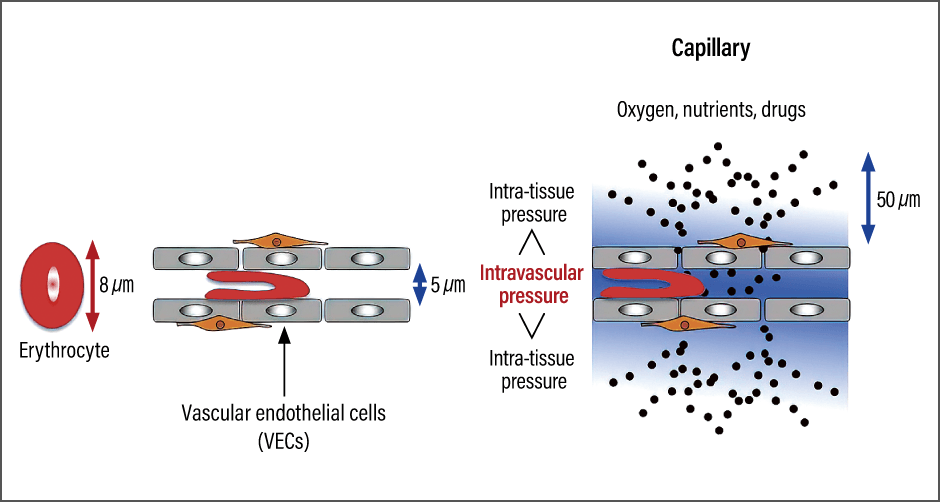
Figure 1. How oxygen, nutrients, and drugs are transportedLarger in diameter than the internal diameter of a capillary, the disc-shaped erythrocyte folds in on itself to pass through the blood vessel. When it does so, oxygen, nutrients, and drugs are pushed out from the blood vessel and are delivered to areas with lower tissue pressure.
Another important thing is the distance between capillaries. Capillaries are spaced out at more or less regular intervals, locating neatly in parallel. Transported by differences in osmotic pressure, oxygen and nutrients can reach a distance of as much as 50 µm from the capillary. Accordingly, capillaries are spaced out at intervals of 100 µm (50 µm + 50 µm), to ensure the supply of oxygen and nutrients to tissues. However, in skeletal muscle, which has a high oxygen demand, the distance between capillaries is smaller.
Looking at the fingernail using a blood flow microscope, i.e., dermal microscopy, one can see that the capillaries in people with healthy tissue run neatly in parallel with each other. However, they are sparser in aged tissue. This is because the capillaries become thinner and the blood flow through them declines. I have named these thinner capillaries “ghost vessels” because those have not been designated thus far. Ghost vessels cannot carry oxygen or nutrients to the surrounding tissue. Consequently, the tissue progressively becomes damaged and its functions decline.
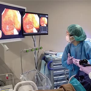
I began researching ghost vessels based on the inefficacious of anti-cancer drug for cancer patients when I was a clinician working in a department of hematology and oncology. I wondered whether there might be a problem with the drug delivery. The anti-cancer drug administered was delivered to cancer cells via the capillaries. When I investigated the cancer capillaries, I found that many of those inside the cancer tissue were immature and unable to perform their proper function, which meant neither oxygen nor the anti-cancer drug were able to be delivered to the tissue.
When oxygen is not being transported
The photograph in Figure 2 shows colon cancer tissue; red indicates capillaries, green indicates hypoxia, and blue indicates cancer cells penetrated by the drug (a DNA dye, in this case). From this photograph, we can see that although there are numerous capillaries in the cancer tissue, oxygen is not transported to them.
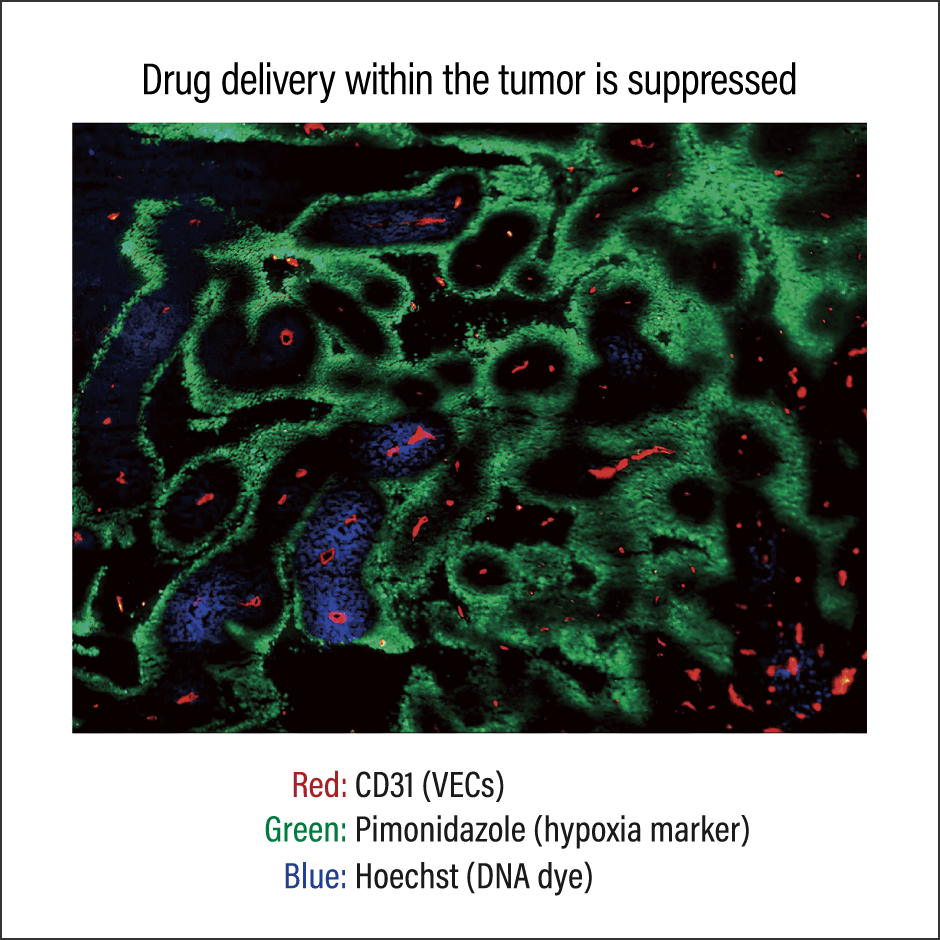
Figure 2. Photograph of colon cancer tissueEven though the cancer tissue has capillaries, delivery of oxygen and drugs is insufficient. Due to the hypoxic environment, immune cells cannot function.
Even if the leukocytes (immune cells) that destroy cancer cells enter this cancer tissue, they are unable to fulfill their immune function, because immune cells are highly vulnerable to hypoxia.
Cancer cells, on the other hand, can survive even in comparatively low oxygen environments. They have acquired the ability to produce a high-energy compound called adenosine triphosphate (ATP) without using oxygen, in a process called anaerobic glycolysis. Accordingly, cancer cells can live quite freely as long as they avoid attack by lymphocytes. To put it another way, cancer cells could be said to deliberately create a hypoxic environment in order to avoid attack by lymphocytes.
The reality of whether cancer cells can be destroyed with the aid of anti-cancer drugs is that the drugs only enter some of the cancer cells, so, administering anti-cancer drugs does not always work. I spent around 20 years analyzing why drugs do not enter the parenchyma of tumor.
Mural cells called pericytes are arranged at regular intervals around the VECs of the capillaries. Precise observation of the capillaries in cancer tissue reveals that they have very few of these mural cells, with no cells binding the capillaries together from the outside. This causes blood vessels to become distended, with a deformed structure. Gaps between VECs also become wider, making them more prone to leakage, while the ends of the capillaries narrow —— this is the process of ghost vessel formation (Figure 3).
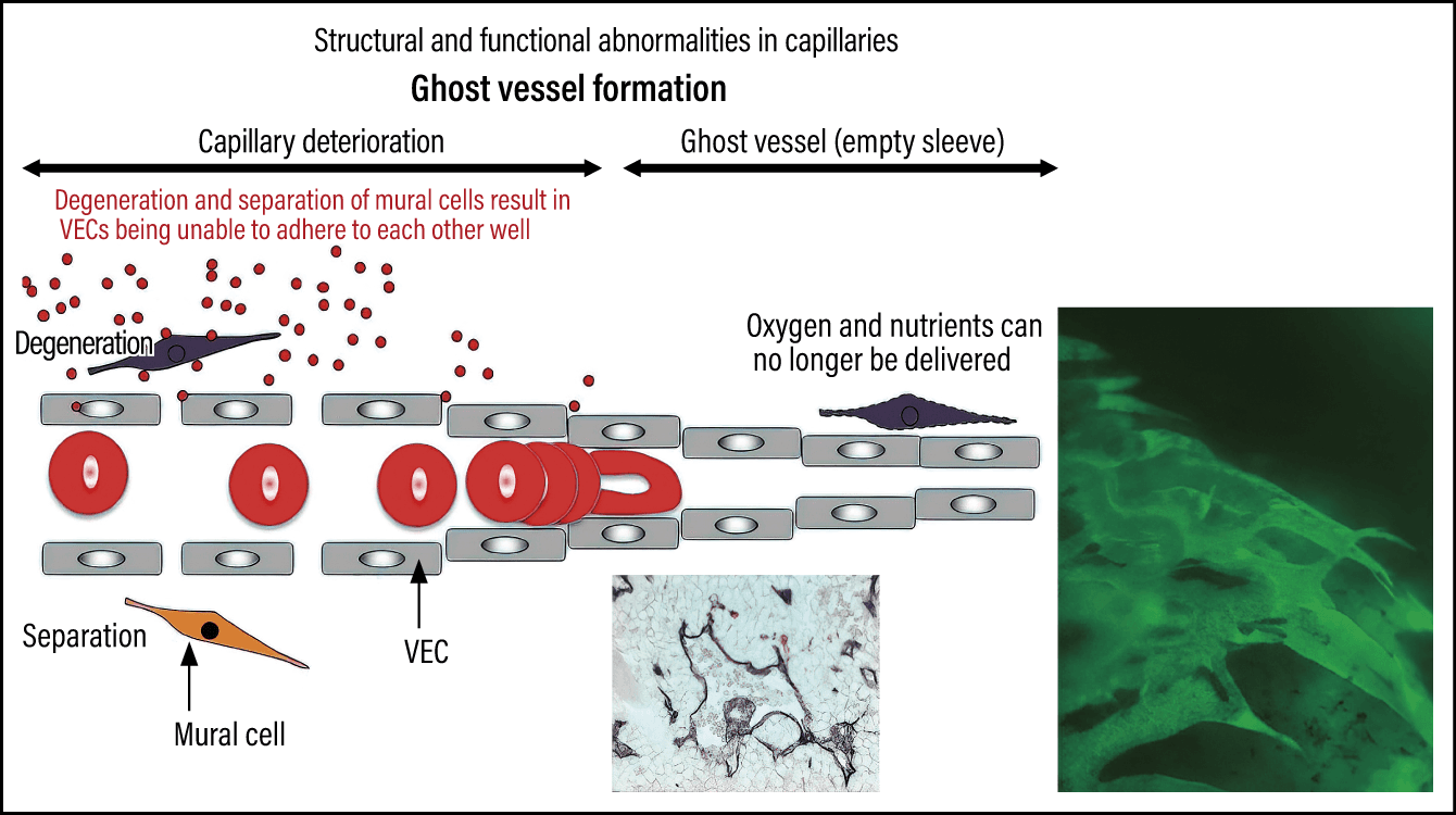
Figure 3. The mechanism of ghost vessel formationIf the pericytes that adhere around the outside of VECs separate from them, the blood vessel dilates and becomes deformed, causing the capillaries at the tip to narrow. As a result, blood serum builds up, preventing oxygen, nutrients, and drugs from reaching the tissue.
It has been discovered that the formation of ghost vessels in this way in healthy human bodies can trigger a variety of conditions, including lifestyle-related diseases.
The sparse capillaries of elderly people
One such condition is skin aging. If we compare the skin of a six-year-old with that of someone aged 75, we can see that, whereas the child’s capillaries are located at 100 µm intervals, the elderly person has only one capillary every few hundred micrometers. In this state, oxygen and nutrients do not reach all of the tissue and the skin gradually becomes thinner as dermal collagen production is suppressed, causing wrinkles and other signs of skin aging. The mechanism of skin aging in association with capillaries was first reported 40 years ago.
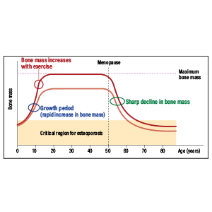
Osteoporosis is said to affect more than 10 million people, most of them elderly. Inside our bones is a spongy form of bone tissue called trabecular bone, which transfers the load of our weight. Osteoporosis is a condition in which this spongy bone tissue gradually becomes brittle.
Bone is able to regenerate thanks to nutrients delivered by capillaries running through fine trabecular bone. However, the number of these capillaries progressively decreases as we age. In 2014, a research team in Germany reported that one of the causes of osteoporosis was likely to be the decline in trabecular bone capillaries that accompanies aging.
It has recently started to become apparent that ghost vessel formation is also a cause of dementia. The brain has highly developed capillary networks, but scientists have discovered from experiments in mice that the number of capillaries in the cerebral parenchyma declines with age.
Observation of cerebral capillaries in patients with Alzheimer’s disease and in healthy individuals reveals that, as a protein called amyloid builds up in the brain of Alzheimer’s patients, the number of capillaries falls to between two-thirds and half of the number found in healthy individuals. The current hot topic in this field of research is the possibility that amyloid —— a waste product —— might build up because the decrease in capillaries means it is not discharged, and that this accumulation might be the cause of dementia.
Age-related macular degeneration (AMD) is an eye disorder in which the macula, which is located in the center of the retina, is damaged by hemorrhage and edema as we age, causing vision impairment. It is becoming clear that this, too, is caused by an increase in immature capillaries.
These conditions and lifestyle-related diseases occur as we age because of problems in our lifestyles. Along with excessive intake of fat and salt, hypertension causes mural cells to degenerate, and there is said to be a particularly close relationship to elevated blood glucose levels.
A high blood glucose level guides reactive oxygen species (ROS) to VECs and mural cells. When mural cells are damaged by degeneration caused by these ROS, gaps become wider between VECs, causing oxygen and nutrients to leak out. It is said that this is what causes people’s tissue to swell, and if this progresses, ghost vessels are formed.
When the blood glucose level rises, the excess sugar binds with proteins to form advanced glycation end products (AGEs). When that occurs, the mural cells of the capillaries progressively degenerate and break away. It has been discovered that this causes organ function to decline, which leads to aging when it occurs throughout the body.
Until now, research into vascular aging had focused primarily on arteriosclerosis. It is conceivable that the decline in the number of capillaries resulting from the formation of ghost vessels is quite closely related to actual tissue and organ aging. In fact, as long ago as 1963, a U.S. research team reported on the relationship between diabetes and mural cells.
Improving capillaries at risk of becoming ghost vessels
So, how can we prevent capillaries from becoming ghost vessels?
First and foremost, paying attention to our blood glucose levels is crucial. Even if our ordinary and fasting blood glucose levels are normal, eating a lot of carbohydrates such as rice and sweet things all at once or drinking large quantities of sugary beverages causes blood glucose to shoot up. These transient sharp rises in blood glucose levels are called glucose spikes and could produce capillaries more prone to forming ghost vessels. Take care to consume a good balance of nutrients and not to overeat.
Moderate exercise is effective in improving capillaries at risk of becoming ghost vessels. Taking moderate exercise improves blood flow, encouraging the expression of vascular cell adhesion molecules, which help VECs to adhere to each other. Around 20-30 minutes of walking brisk enough to work up a light sweat is fine.
In daily life, stress and lack of sleep are bad for your blood vessels. When you are tense, your sympathetic nervous system takes priority and the arterioles that feed the capillaries constrict, reducing blood flow. To improve blood flow, you need to relax, so that the parasympathetic nervous system can take priority, and also get plenty of sleep. High-quality sleep promotes the overnight secretion of hormones that repair blood vessels. You can also relax by soaking in a bath of warm water (a temperature of about 40°C is ideal).
When mural cells produce a substance called angiopoietin-1, a receptor called Tie2 is activated, maintaining the blood vessel in a comparatively stable state (Figure 4). In fact, in March 2022, approval was granted for a drug that activates Tie2 to be indicated in cases of AMD and diabetic retinopathy and the drug began to be administered.
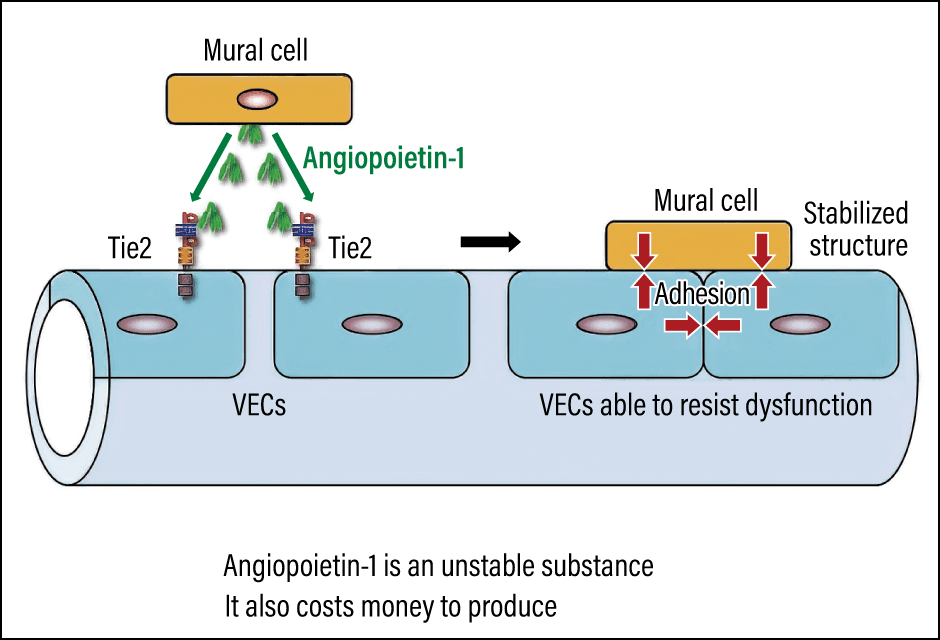
Figure 4. Stabilization of vascular structure by promoting Tie2 activationReceptors called Tie2 are involved in the adhesion of VECs and mural cells. When mural cells produce a substance called angiopoietin-1, Tie2 is activated, enabling the blood vessel to be maintained in a comparatively stable state.
However, the drug cannot be used to address the risk of developing ghost vessels. Accordingly, we are focusing on controlling the receptors involved in the adhesion of mural cells and Tie2. Investigating whether there are any substances in food capable of activating Tie2 receptors, we found three that demonstrated effects: beta-syringaresinol in cinnamon, a type of flavonoid among the polyphenols in rooibos tea, and piperine in long pepper, a spice widely used in Okinawa (Figure 5).
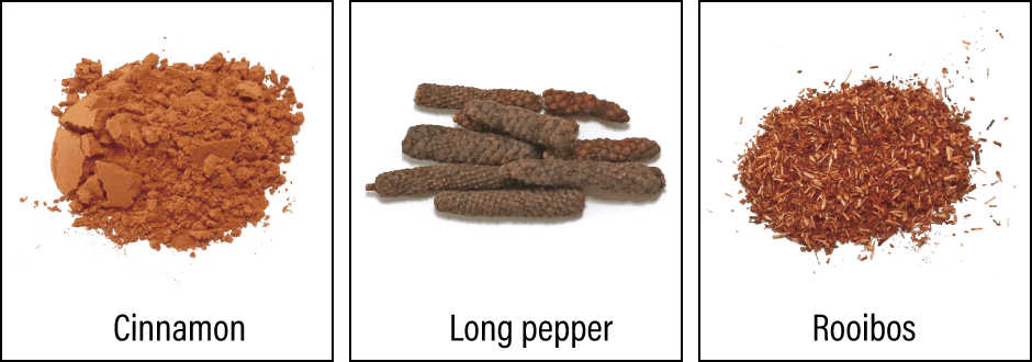
Figure 5. Substances capable of activating Tie2 receptorsSubstances found in cinnamon, rooibos, and long pepper were found to be effective in activating Tie2 receptors.
Most medical facilities currently provide exercise therapy and anti-cancer drugs. Some facilities overseas provide therapies that combine yoga with anti-cancer drugs. There have even been reports providing data to show that light stretching while in a relaxed state increased the effectiveness of anti-cancer drugs by cleansing the blood vessels.
I believe that restoring the normal functioning of capillaries is crucial to treating cancer.
(Figures courtesy of Nobuyuki Takakura)